导语
EVO Toric ICL(TICL)矫正近视散光是安全、有效且可预测的,但TICL的精准性和旋转稳定性仍然值得关注1-5。术后拱高较低被认为是TICL旋转的潜在风险,但这一假设尚未得到大样本临床研究的证实。也有人认为脚襻位置不当可能会影响TICL的旋转稳定性4,13。本研究评估了TICL术后低拱高眼的临床结果和旋转稳定性,并研究脚襻位置和低拱高(拱高<250μm)是否会影响TICL的旋转稳定性。
研究结果由中山大学中山眼科中心的余克明教授团队于2024年7月发表在Journal of Refractive Surgery杂志上。
研究方法
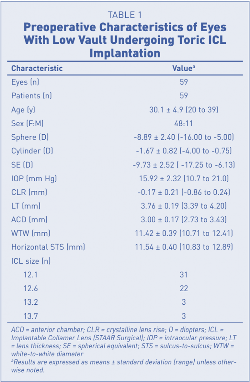
本研究纳入2020年10月至2022年10月接受TICL植入术的1017名患者中的59名患者的59只术后低拱高眼(从TICL术后一周到末次随访期间拱高始终低于250μm),平均随访六个月。检查项目包括:裸眼远视力(UDVA)、矫正远视力(CDVA)、屈光度、眼压(IOP)、拱高、UBM检查以及裂隙灯检查。
EVO ICL术后脚襻位置进一步定义为以下四种类型12:(1)脚襻插入睫状沟;(2)脚襻在睫状突上方;(3)脚襻插入睫状突;(4)脚襻在睫状突下方。
研究结果
共纳入59名患者的59只眼睛,平均年龄为30.1±4.9岁(范围:20至39岁),术前平均SE为-9.73±2.52D(范围:-17.25至-6.13D)(表1)。所有手术均顺利完成,无术中并发症,所有患者均观察6个月或更长时间。随访期间,未出现其他并发症,包括前囊下白内障、眼压升高等。术后六个月,平均中央拱高为137.4±61.0µm(范围:40至236µm),19只眼睛的术后拱高≤100µm。
视力及眼压结果
术后6个月,88%(52/59)的眼睛UDVA达到20/20或更好,98%的眼睛UDVA达到20/25或更好。平均有效性指数和安全性指数分别为1.04和1.15。末次随访时,没有患者出现CDVA丢失(图1)。
平均MRSE从术前的-9.73±2.52D显著下降至术后6个月的-0.01±0.33D。93%和100%的眼睛在目标SE的±0.50和±1.00D范围内。平均显然验光散光度数从术前的-1.67±0.82D改善到术后6个月的-0.43±0.33D。50(84.7%)和57(96.6%)眼的术后散光度数分别在±0.50和±1.00D内,表明TICL对于矫正低拱高眼的近视散光具有良好的疗效和可预测性。表A和图A显示了术后6个月散光矢量分析的结果。
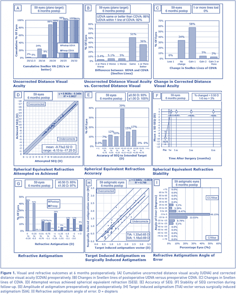
(图1)
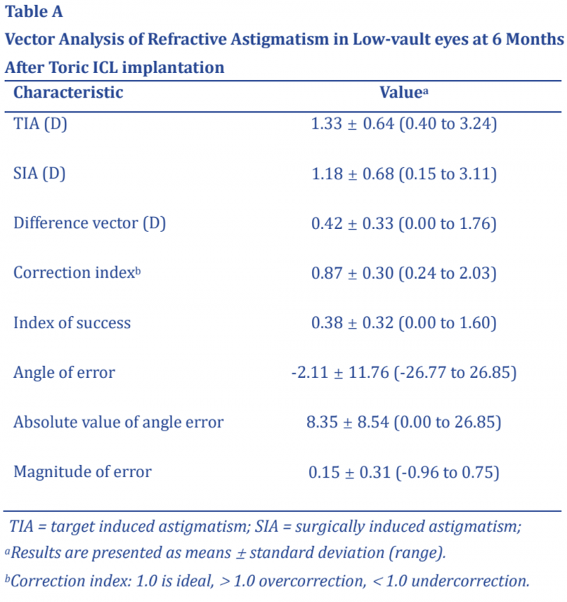
(表A)
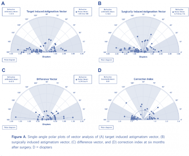
(图A)
旋转稳定性和脚襻位置
本研究中,术中TICL放置角度与水平轴位差值的平均值为5.05±4.00°(范围:0至15.00°)。术后1周、1个月、3个月和6个月时,TICL旋转度数(指TICL术后实际角度与术中放置角度差值的绝对值)平均值分别为3.98±3.03°(范围:0至11.40°)、4.30±2.98°(范围:0至13.03°)、4.23±3.17°(范围:0至12.70°)和4.50±3.08°(范围:0至12.50°)。从术后一周到末次随访,旋转度数没有明显差异。术后6个月,35.6%的眼睛(21/59)旋转度数≤3°,61.0%的眼睛(36/59)旋转度数≤5°,只有5只眼睛旋转度数≥10°。没有眼睛需要调位。
表2显示了ICL脚襻的术后位置。共有28.8%(17/59)的眼睛所有脚襻位于睫状沟或睫状突上方。33.9%(20)的眼睛3个脚襻位于睫状突上方,25.4%(15)的眼睛2个脚襻位于睫状沟或睫状突上方,4只眼只有1个脚襻位于睫状突上方,3只眼的4个脚襻均在睫状突下方。

与旋转相关的危险因素
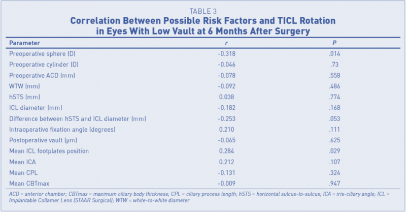
如表3所示,TICL旋转度数与术前球镜度数(r=-0.318,P=.014)以及脚襻位置平均值(r=0.284,P=.029)之间存在显著相关性。TICL旋转度数与术后散光残留度数呈正相关(r=-.469,P≤ .001)。
讨论
本研究术后平均拱高为137.4±61.0µm,分别有76.3%和32.2%的眼睛低于200µm和100µm。术后6个月时,有效性和安全性指数分别为1.05和1.16,与之前研究的结果一致3,14-16。55只眼睛(93.2%)术后SE在目标SE的±0.50D以内。表明TICL在低拱高眼中具有良好的近视散光矫正效果。
术后六个月有50(84.7%)和57(96.6%)只眼睛散光在±0.50和±1.00D内,与Zhao等人15和Lee等人14报道的正常拱高眼的散光结果相似。只有4只眼术后散光≥1.00D(范围:1.00至1.75D),平均旋转8.82°(范围:7.59至12.02°),其中3眼术后UDVA均等于或优于术前CDVA。
术后六个月的TICL旋转度数为4.50±3.08°,与先前报道结果相当3,14,19。所有TICL的旋转度数均小于15°,只有5只眼睛旋转超过10°,其UDVA均为20/20,残余散光均≤1.00D。总而言之,TICL低拱高眼的视觉结果和旋转稳定性基本与拱高正常的眼睛相同。
本研究发现旋转度数和脚襻位置之间存在显著相关性。当脚襻位于睫状沟内时,TICL似乎更稳定,与Park等人的结果一致4,与Sheng等人的结果相反13。理论上,当脚襻位于睫状沟内时,脚襻末端被睫状突和虹膜固定。当脚襻插入或低于睫状突时,其位置不正可能引起TICL的压力传递异常,这可能会导致TICL倾斜或松动,最终增加旋转的风险。
在本研究中,术后拱高与TICL旋转角度之间没有观察到显著相关性,与之前研究结果一致3,4,8,14,22。多种因素可能导致拱高异常,包括ICL尺寸不合适、自然晶状体厚且前表面矢高高以及ICL脚襻错位。本研究中,有19只眼睛术后拱高小于100µm,只有一只眼睛的旋转度数大于10°。因此,相比于自然晶状体因素导致的低拱高,由于脚襻错位或ICL尺寸不合适而导致的低拱高更有可能与术后旋转相关。
TAKE HOME MESSAGE
1、EVO TICL在低拱高眼中具有良好的有效性、安全性、可预测性,且旋转稳定性较好,基本与正常拱高的眼睛相同。
2、EVO TICL的旋转稳定性与脚襻位置相关,提示应关注术后低拱高患者的脚襻位置。
文献来源
https://pubmed.ncbi.nlm.nih.gov/39007814/
参考文献
1. Alfonso JF, Lisa C, Alfonso-Bartolozzi B, Pérez-Vives C, Montés-Micó R. Collagen copolymer toric phakic intraocular lens for myopic astigmatism: one-year follow-up. J Cataract Refract Surg. 2014;40(7):1155-1162. https://doi.org/10.1016/j.jcrs.2013.11.034 PMID:24957435
2. Hashem AN, El Danasoury AM, Anwar HM. Axis alignment and rotational stability after implantation of the toric implantable collamer lens for myopic astigmatism. J Refract Surg. 2009;25(10)(suppl):S939-S943.https://doi.org/10.3928/1081597X-20090915-08 PMID:19848375
3. Hyun J, Lim DH, Eo DR, Hwang S, Chung ES, Chung TY. A comparison of visual outcome and rotational stability of two types of toric implantable collamer lenses (TICL): V4 versus V4c. PLoS One. 2017;12(8):e0183335. https://doi.org/10.1371/journal.pone.0183335 PMID:28846701
4. Park SC, Kwun YK, Chung ES, Ahn K, Chung TY. Postoperative astigmatism and axis stability after implantation of the STAAR Toric Implantable Collamer Lens. J Refract Surg. 2009;25(5):403-409. https://doi.org/10.3928/1081597X-20090422-01 PMID:19507791
5. Sanders DR, Schneider D, Martin R, et al. Toric Implantable Collamer Lens for moderate to high myopic astigmatism. Ophthalmology. 2007;114(1):54-61. https://doi.org/10.1016/j.ophtha.2006.08.049 PMID:17198849
6. Titiyal JS, Kaur M, Jose CP, Falera R, Kinkar A, Bageshwar LM. Comparative evaluation of toric intraocular lens alignment and visual quality with image-guided surgery and conventional three-step manual marking. Clin Ophthalmol. 2018;12:747- 753. https://doi.org/10.2147/OPTH.S164175 PMID:29731603
7. Schartmüller D, Schriefl S, Schwarzenbacher L, Leydolt C, Menapace R. True rotational stability of a single-piece hydrophobic intraocular lens. Br J Ophthalmol. 2019;103(2):186-190. https:// doi.org/10.1136/bjophthalmol-2017-311797 PMID:29666120
8. Mori T, Yokoyama S, Kojima T, et al. Factors affecting rotation of a posterior chamber collagen copolymer toric phakic intraocular lens. J Cataract Refract Surg. 2012;38(4):568-573. https://doi.org/10.1016/j.jcrs.2011.11.028 PMID:22342008
9. Reinstein DZ, Vida RS, Archer TJ. Visual outcomes, footplate position and vault achieved with the Visian implantable collamer lens for myopic astigmatism. Clin Ophthalmol. 2021;15:4485- 4497. https://doi.org/10.2147/OPTH.S330879 PMID:34848942
10. Shi M, Kong J, Li X, Yan Q, Zhang J. Observing implantable collamer lens dislocation by panoramic ultrasound biomicroscopy. Eye (Lond). 2015;29(4):499-504. https://doi.org/10.1038/ eye.2014.336 PMID:25613840
11. Zhang X, Chen X, Wang X, Yuan F, Zhou X. Analysis of intraocular positions of posterior implantable collamer lens by full-scale ultrasound biomicroscopy. BMC Ophthalmol. 2018;18(1):114. https://doi.org/10.1186/s12886-018-0783-5 PMID:29743110
12. Yiming Y, Xi C, Huan Y, et al. Evaluation of ciliary body morphology and position of the implantable collamer lens in low-vault eyes using ultrasound biomicroscopy. J Cataract Refract Surg. 2023;49(11):1133-1139. https://doi.org/10.1097/j. jcrs.0000000000001285 PMID:37586102
13. Sheng XL, Rong WN, Jia Q, et al. Outcomes and possible risk factors associated with axis alignment and rotational stability after implantation of the Toric implantable collamer lens for high myopic astigmatism. Int J Ophthalmol. 2012;5(4):459-465. PMID:22937505
14. Lee H, Kang DSY, Choi JY, et al. Rotational stability and visual outcomes of V4c toric phakic intraocular lenses. J Refract Surg. 2018;34(7):489-496. https://doi.org/10.3928/108159 7X-20180521-01 PMID:30001453
15. Zhao J, Zhao J, Yang W, et al. Consecutive contralateral comparison of toric and non-toric implantable collamer lenses V4c in vault after implantation for myopia and astigmatism. Acta Ophthalmol. 2021;99(6):e852-e859. https://doi.org/10.1111/ aos.14720 PMID:33369209
16. Nie D, Yan P, Yan Z, et al. Polar value analysis of astigmatic change and rotational stability after implantation of V4c toric implantable collamer lens. Ann Transl Med. 2021;9(2):139. https://doi.org/10.21037/atm-20-7835 PMID:33569441
17. Felipe A, Artigas JM, Díez-Ajenjo A, García-Domene C, Alcocer P. Residual astigmatism produced by toric intraocular lens rotation. J Cataract Refract Surg. 2011;37(10):1895-1901. https:// doi.org/10.1016/j.jcrs.2011.04.036 PMID:21865007
18. Potvin R, Kramer BA, Hardten DR, Berdahl JP. Toric intraocular lens orientation and residual refractive astigmatism: an analysis. Clin Ophthalmol. 2016;10:1829-1836. https://doi. org/10.2147/OPTH.S114118 PMID:27703323
19. Wei PH, Li J, Jiao XL, Yu Z, Song H. Short-term clinic observation of misalignment and rotational stability after implantable collamer lens implantation. Graefes Arch Clin Exp Ophthalmol. 2023;261(5):1473-1481. https://doi.org/10.1007/s00417- 022-05929-7 PMID:36484805
20. Chen Q, Tan W, Lei X, et al. Clinical prediction of excessive vault after implantable collamer lens implantation using ciliary body morphology. J Refract Surg. 2020;36(6):380-387. https:// doi.org/10.3928/1081597X-20200513-02 PMID:32521025
21. García-Feijoó J, Alfaro IJ, Cuiña-Sardiña R, Méndez-Hernandez C, Del Castillo JM, García-Sánchez J. Ultrasound biomicroscopy examination of posterior chamber phakic intraocular lens position. Ophthalmology. 2003;110(1):163-172. https://doi. org/10.1016/S0161-6420(02)01449-5 PMID:12511362
22. Zhu M, Zhu L, Zhu Q, et al. Clinical effect and rotational stability of TICL in the treatment of myopic astigmatism. J Ophthalmol. 2020;2020:3095302. https://doi.org/10.1155/2020/3095302 PMID:3348932


